
“A Model for the Interfacial Kinetics of Phospholipase D Activity on Long-Chain Lipids”
Majd S., Yusko E. C., Yang J., Sept D.*, and Mayer M.*
Biophysical Journal, 2013, 105, 146-153.
The cover image illustrates the enzymatic activity of phospholipase D (PLD), a soluble enzyme that hydrolyzes phospholipids, at the membrane interface. This image, composed of an Adobe Illustrator cartoon and a colorful image of a night sky, was prepared by Sheereen Majd and reflects the focus of our study on PLD enzymatic kinetics. These kinetics are complex as the lipid products of this reaction are charged and remain in the membrane, affecting the enzyme-membrane binding kinetics and hence, the overall enzymatic kinetics. As illustrated, the accumulation of the charged lipids in the membrane can be probed locally by changes in ion conductance of gramicidin channels. In this study, we used the channel conductance measurements to derive the kinetics of PLD catalytic reaction and developed a model that describes these kinetics by considering the effect of product accumulation and substrate depletion in the membrane.
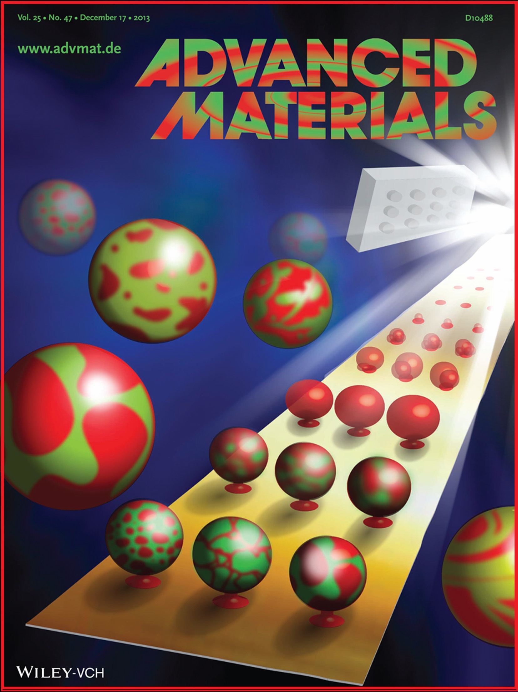
“A Simple and Versatile Method for the Formation of Arrays of Giant Vesicles with Controlled Size and Composition”
Kang Y. J., Wostein H. S., and Majd S.*
Advanced Materials, 2013, 25, 6834-6838.
This cover image illustrates the concept of the presented approach for the preparation of cell-sized liposomes with functional membrane proteins. Liposomes prepared in size scale of cells and with lipid and protein components similar to those found in natural cell membranes are referred to as giant proteoliposomes and closely mimic cellular membranes. As demonstrated in this cartoon, prepared by You Jung Kang, this article introduces a unique, low-cost, and versatile approach for producing uniformly sized giant proteoliposomes. In this approach, a hydrogel stamp deposits lipids and proteins on a substrate. These lipid/protein deposits are next exposed to an AC electric field that leads to the formation of giant proteoliposomes of controlled size and composition. This method of production of giant proteoliposomes can be useful in biomaterials, biotechnology, and biosensing applications as well as in the biophysical fundamental studies.
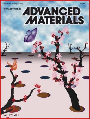
Park S., Yang G., Madduri N., Abidian M. R., and Majd S.*
Advanced Materials, 2014, 26, 2782–2787.
The cover image illustrates conducting polymer films, grown in a patterned fashion, that are decorated with variety of biomolecules such as antibodies or proteins (represented by the flowers) to attract cells or other biomolecules (represented by the butterflies). This artistic image, created by SooHyun Park, represents the focus of this article on generating patterned films of conducting polymers with various geometries and surface chemistries using a novel and simple methodology. This method, referred to as hydrogel-mediated electropolymerization, employs hydrogel stamps as carrier for polymer precursors and enables direct micro-patterning of conducting polymers with multiple chemistries and bioactive molecules in a single-step and solution-free process. These patterned films of bio-functionalized conducting polymers are attractive platforms for biomedical applications such as biosensing and cell/tissue engineering.
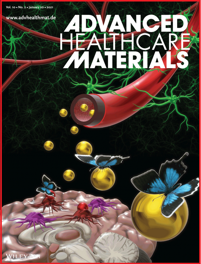
“Tumor Targeted Delivery of an Anti‐Cancer Therapeutic: An In Vitro and In Vivo Evaluation”
Kang Y.J., Holley C.K., Abidian M., Madhankumar A.B., Connor J., and Majd S.*
[Kang Y.J. and Holley C.K. contributed equally]
Advanced Healthcare Materials, 2021, 10, 2001261
The cover image presents the focus of this article on a new chelator-based nano-formulation for treatment of highly aggressive glioma tumors. In this study, biodegradable nanoparticles that are loaded with a potent anti-cancer iron chelator, Dp44mT, and decorated with a glioma tumor-targeting ligand, IL13, are developed and their effect on the growth of glioma tumors is evaluated in vitro and in vivo. In this artistic illustration, prepared by Dr. You Jung Kang, IL13 ligands (represented by butterflies) carry the Dp44mT-loaded nanoparticles (represented by honey) from the vein (represented by the nectarine of a flower) to the tumor (represented by the nest), feeding and destroying the tumor. This work is the first to demonstrate the great potential of targeted delivery of Dp44mT for cancer treatment.